JI Core Service Request Form
The goal of the Joint Imaging (JI) Core is to provide MARC members with access to magnetic resonance imaging (MRI) of human and animal joints.
What can we learn from the MRI of joints? Depending on the technique used, you can learn about basic anatomy, tissue pathology, and treatment effects.
Who and what can we image? In clinical scanners (3-Tesla), we can image all major body parts in live human subjects, along with large-to-small animal specimens. We utilized radio-frequency coils of varying sizes (can fit diameters of 1" to ~18") to acquire the best image possible for each case. In preclinical scanners (3- to 11-Tesla), we can image small live animals (mice and rats) and specimens.
MRI Techniques for the Knee (#: available on human scanner only)
- proton density weighted (PD): anatomy to distinguish joint fluid, cartilage, menisci, and ligaments
- PD or T2 fat suppressed: meniscal tear detection, cysts, and edema of bone marrow and fat pad
- T1 weighted: bone structure and soft tissue lesion
-
# ultrashort echo time (UTE): Can be used in lieu of CT to image bone by assessing intrasubstance structure of short T2 tissues including the osteochondral junction, menisci, and ligaments

- T2 mapping: correlates with water content and collagen fibril organization in articular cartilage and meniscus, increases with degeneration
- T1rho mapping: correlates with water and proteoglycan content in cartilage, also increases with degeneration
- # UTE T2* mapping: similar to T2 but stronger correlation with degeneration and pathology

Examples on rat and mouse knees:

- T2, T1rho, T2* color mapping
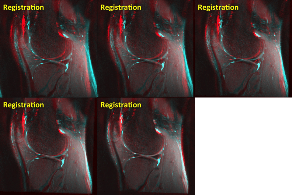
- Image registration

3. 3D rendering