Overview
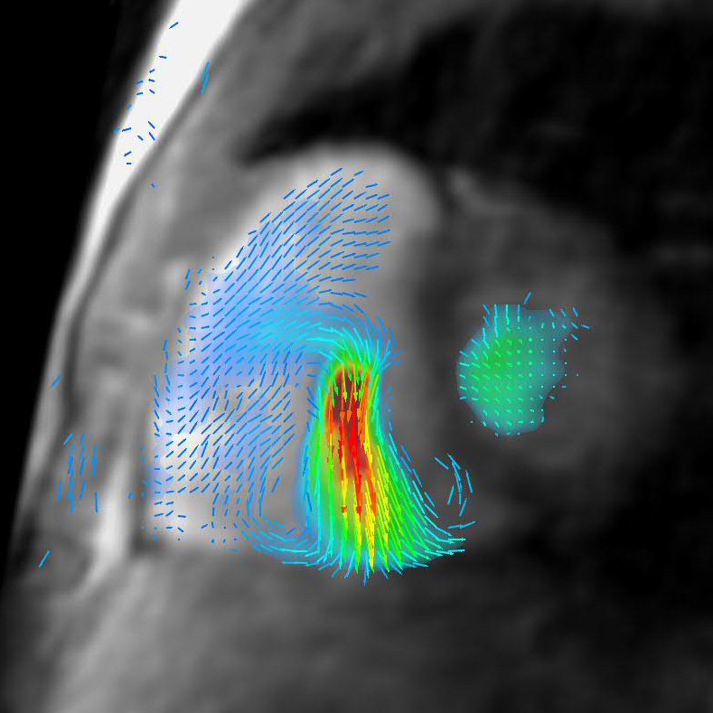
CTIPM’s cardiovascular imaging lab is developing and applying advanced MRI techniques to the diagnosis of cardiovascular disease, specifically the use of 4D Flow MRI to study the heart and blood vessels throughout the body. Clinical-translational MRI scans performed by our laboratory are designed to maximize patient comfort, while obtaining all of the relevant clinical parameters in the least amount of time (usually 30-45 minutes). The lab is focused on applying leading edge advanced imaging techniques to immediately diagnose and manage cardiovascular diseases today, while also developing the strategies for tomorrow. We have an ongoing partnership with Arterys to develop the latest cloud-based computational technologies for advanced visualization and diagnosis. Research focus areas include MRI Characterization of Cardiovascular Fluid Mechanics, Translational Computational Cardiovascular Biomechanics, and Multimodality Integrated Cardiac Imaging.
Clinical Applications
Indications
- Atrial septal defect
- Ventricular septal defect
- Tetralogy of Fallot
- Aortic coarctation
- Partial anomalous pulmonary venous return
- Single ventricle – Glenn, Fontan
- Other complex congenital heart conditions
Common Components
- MRA Pulmonary and/or Aortic Arteriogram
- Right Ventricular Volumetry, Function
- Quantification of Pulmonary, Tricuspid Regurgitant Fraction
- Quantification of Pulmonary Stenosis Pressure Gradient
- Quantification of Split Pulmonary Perfusion
- Quantification of Intracardiac, Extracardiac Shunts, Shunt Fraction


Indications
- Aortic stenosis, regurgitation
- Mitral stenosis, regurgitation
Common Components
- Left Ventricular Volumetry, Function
- Quantification of Aortic Stenosis, Regurgitant Fraction
- Quantification of Mitral Stenosis, Regurgitant Fraction
|
| Regurgitant fraction 60% (severe), requiring repeat valve surgery. |
Indications
- Pulmonary hypertension
- Chronic thromboembolic disease
Common Components
- MRA Pulmonary Arteriogram
- Right Ventricular Volumetry, Function
- Estimation of Right Ventricular Systolic Pressure
- Quantification of Pulmonary, Tricuspid Regurgitant Fraction
- Quantification of Split Pulmonary Perfusion
Indications
- Ascending aortic aneurysm (+/- bicuspid aortic valve)
- Aortic dissection
- Peripheral arterial disease
Common Components
- MRA Arteriogram
- Longitudinal follow-up of aortic dimensions
- Quantification of arterial stenosis

| 
|
|
Indications
- Deep venous thrombosis
- May-Thurner syndrome
- Central venous stenosis
Common Components
- MRA Venogram
- Quantification of venous stenosis
- Quantification of venous collateral flow
Indications
- Cervical, thoracic, abdominal vasculitis
Common Components
- MRA Arteriogram
- Characterization of wall thickening, edema, enhancement
Indications
- Fibroids
- Pelvic congestion
Common Components
- Mapping of uterine and ovarian arteries
- Mapping and quantification of ovarian venous flow

Director:
 Albert Hsiao, MD/PhD
Assistant Professor of Radiology at UC San Diego
Director of Cardiovascular Imaging, CTIPM
Affiliated Clinical Services: