Prostate cancer is the most common cancer among American men. While most prostate cancers grow slowly and don’t cause any health problems, the disease still kills more than 27,000 people in the United States annually. For physicians and their patients, it’s critical to quickly detect, localize and characterize tumors through advanced imaging and timely biopsies of the prostate.
UC San Diego Health is a national leader in the use of magnetic resonance imaging (MRI) technology to guide the diagnosis and treatment of various cancers, including brain and prostate malignancies. UC San Diego oncologists affiliated with the Center for Translational Imaging and Precision Medicine (CTIPM) have advanced the diagnosis of prostate cancer on several fronts.
Combining MRI with ultrasound imaging for tumor detection and more targeted biospies
In one innovation, physicians have combined magnetic resonance imaging (MRI) technology with a traditional ultrasound prostate exam to generate a three-dimensional map of the prostate with high tumor contrast. Ultrasounds offer only imperfect views of the prostate, and tumors can be missed. But MR imaging enhances what physicians can see, leading to a more precise detection of cancer, and better-targeted biopsies of prostate tissue.
The experience of Armondo Lopez, a recent patient at Moores Cancer Center at UC San Diego, illustrates the power of combining imaging techniques. Mr. Lopez had been given a clean bill of health after physicians had performed a traditional biopsy guided by ultrasound imaging. But when levels of his prostate-specific antigen (PSA), a protein often elevated in men with prostate cancer, started to rise he began to worry. His physician at UC San Diego recommended an MRI-guided prostate biopsy. The new technology led to the diagnosis of an aggressive prostate cancer situated in an area of the gland that is difficult to biopsy. Fortunately for Mr. Lopez, the tumor was still in an early stage and treatable. An early diagnosis typically improves a patient’s prognosis.
For patients, the only added step to their prostate examination is the addition of an MRI, which occurs during a separate visit in advance of the biopsy exam. The MRI data are combined with real-time, ultrasound-guided biopsy images in the clinic, offering the physician conducting the biopsy a 3D road map of the prostate.

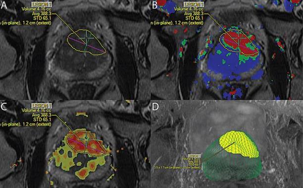
MRI technology was used to identify and locate a probable tumor (outlined in yellow) during a targeted prostate biopsy for a patient who had previously had multiple negative biopsies but had persistently high PSA levels. The biopsy confirmed the presence of high-grade cancer.
A breakthrough advance in MR imaging of the prostate
Another exciting advance in imaging has been pioneered by CTIPM researchers. It’s called restriction spectrum imaging-MRI, or RSI-MRI. It’s one of the latest advances in the practice of quantitative MRI, in which physicians extract objective, quantitative information about the characteristics and progression of disease from magnetic resonance images. Quantitative MRI is also a powerful tool to help guide prostate biopsies.
Dr. David Karow, along with Dr. Anders Dale, Dr. Nathan White and other researchers at UC San Diego, as well as with colleagues at UCLA, first described prostate RSI-MRI in two published papers in early 2015. The new technique allows physicians to more precisely pinpoint a tumor’s location, how extensive it is, and even whether the finding is most likely to represent high or low grade disease – an indication of how quickly a tumor is likely to grow and spread.
Typically for the imaging of tumors, specialists employ contrast enhanced MRI. This involves intravenously injecting die into the patient, which flows to blood vessels feeding the tumor. Because blood flow is commonly enhanced in tumors, physicians can distinguish a tumor from healthy tissue through contrast enhanced MRI. However, this technique can’t detect all tumors because blood flow in some tumors does not differ significantly from blood flow in surrounding tissue.
An alternative technique, known as diffusion MRI, measures the diffusion of water in tissue and therefore the tissue’s density (cancer tumors are generally more dense than healthy tissue). But this technique also has limitations: magnetic field artifacts can distort the actual location of tumors by as much as 1.2 centimeters, or about half an inch. RSI-MRI adjusts for these magnetic field distortions, and also focuses on water diffusion within individual cancer cells – not just through cancer tissue generally. These two improvements allow physicians to more precisely plot a tumor’s location, and its boundaries.
These images illustrate how RSI-MRI data is used for targeted biopsies

Images from the planning software used for targeted biopsy. The tumor is drawn in blue on anatomic imaging overlaid with A) perfusion data and B) RSI-MRI data. The H&E stained histology after prostatectomy D) shows the true boundary of the tumor outline in blue. A 3-D rendering C) of the whole prostate (green) and the tumor (yellow) shows the spatial position of the tumor within the prostate. For this particular patient, RSI-MRI identified a suspicious area, which after biopsy and prostatectomy was proven to be a high-grade tumor. As these figures show, the tumor is barely visible in conventional imaging.

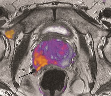
RSI-MRI imaging in the figure to the left shows biopsy proven high-grade cancer. The tumor has extended into the right seminal vesicle (arrow). There is also a region of enhanced RSI signal in the right pelvic bone showing a likely focus of metastatic cancer. The extension of the tumor into the right seminal vesicle, and the likely metastatic bone disease both have implications for subsequent treatment.
What does RSI-MRI mean for the patient? Surely it means a more precise detection and localization of cancerous tumors – through noninvasive imaging and more targeted, precise biopsies. But also, because prostate cancer tumors often can be indolent, a patient may only require active surveillance rather than aggressive treatment. The more advanced RSI-MRI technique offers physicians a powerful way to monitor a patient’s cancer.
Exciting advances in prostate imaging at UC San Diego and its Center for Translational Imaging and Precision Medicine, including these new MR imaging strategies to locate, characterize and biopsy cancerous tumors, continue to make San Diego a national leader in prostate cancer medicine.