Overview
Quantitative MRI is employed to detect, localize and grade tumors within the abdomen and pelvis. Prostate cancer imaging is a specific focus, where non-invasive imaging techniques allow physicians to identify tumor biomarkers. In collaboration with the UC San Diego Department of Urology, the Oncologic Imaging team offers prostate cancer imaging that can detect tumors and guide prostate biopsies.
Images from the planning software used for targeted biopsy

The tumor is drawn in blue on anatomic imaging overlaid with A) perfusion data and B) RSI-MRI data. The H&E stained histology after prostatectomy D) shows the true boundary of the tumor outline in blue. A 3-D rendering C) of the whole prostate (green) and the tumor (yellow) shows the spatial position of the tumor within the prostate. RSI-MRI identified a suspicious area which after biopsy and prostatectomy was proven to be high grade tumor. As shown, the tumor is barely visible in conventional imaging.
Media/Press
Clinical Applications
- Dedicated imaging for screening and staging of abdomen and pelvis tumors
- Screening for prostate cancer
- Dedicated MRI for image-guided prostate biopsies
- Active surveillance of prostate cancer
- Prostate cancer imaging for pre- and post-prostatectomy, radiation therapy and brachytherapy
- Whole body MRI cancer screening
Director
 Michael E. Hahn, MD
Assistant Clinical Professor of Radiology, UC San Diego
Director for Body MRI, CTIPM
Partner Physicians
Department of Urology
Department of Radiation Oncology
Department of Medical Oncology
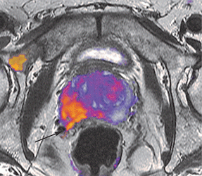
RSI-MRI imaging showing biopsy proven high-grade cancer with extension into the right seminal vesicle (arrow)

There is also a region of enhanced RSI signal in the right pubic bone showing a likely focus of metastatic cancer. The extension into the right seminal vesicle and likely metastatic bone disease has implications for subsequent treatment.